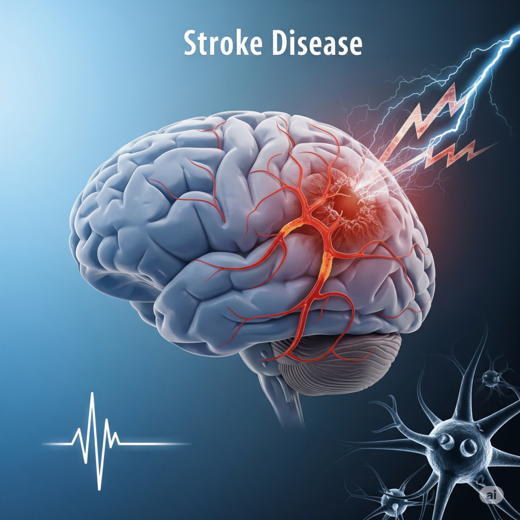
A stroke is a sudden blockage of blood supply in one part of the brain, which prevents brain tissue from obtaining oxygen and nutrients. Within a few minutes, brain cells begin to die, making stroke a medical emergency that requires immediate attention. Depending on the affected brain region, it can cause paralysis, difficulty speaking difficulty, vision problems, loss of balance, or even death. Early diagnosis and treatment are important to reduce brain damage and improve recovery.
It occurs when a blood clot blocks the brain artery, reduces the supply of oxygen. If treated early, it is normal and treatable with clot-busting drugs or mechanical removal.
It occurs when a brain vessel breaks, causing bleeding and loss of pressure. Often associated with high blood pressure or arteriography; Immediate medical care is required.
Lorem ipsum dolor sit amet, consectetur adipiscing elit. Ut elit tellus, luctus nec ullamcorper mattis, pulvinar dapibus leo.
Lorem ipsum dolor sit amet, consectetur adipiscing elit. Ut elit tellus, luctus nec ullamcorper mattis, pulvinar dapibus leo.
Patients may experience sudden weakness, numbness or paralysis on one side of the body, usually affecting the face, arms or legs.
Difficulty may be caused by brain regions controlling brain regions, slide speech, or language.
Sudden blurred vision, dual vision, or complete loss of vision in one or both eyes can be based on the stroke space.
Patients may cause difficulty, dizziness, or loss of balance and coordination, causing movement unstable or unsafe.
A sudden, acute headache – often described as “worst” – indicates a hemorrhagic stroke and requires immediate treatment.
The MRI provides wide brain images to identify stroke space and range. It is particularly useful for detecting small or early strokes remembered on CT scans.
This test uses sound waves to examine narrow or obstructions in carotid arteries. It helps identify atherosclerosis, which can cause ischemic stroke.
A dye is injected into blood vessels, and X-ray images are taken to see blood flow in the brain. It detects obstructions, arterirs or deformities.
Uses ultrasound to create heart images, checks for clotting, valve problems or abnormal structures. Heartmore clots can travel to the brain and cause stroke.
Clotting tests include blood sugar, cholesterol and infection markers. These help to identify the underlying conditions and risk factors that contribute to stroke development.
To detect irregular rhythm such as the atrial fibrillation to measure the electrical activity of the heart, which can cause a leading clot to the stroke.
When the bleeding is suspected, but does not appear on the scan. It detects blood in the cerebrospinal fluid, confirming cerebrospinal hemorrhage.
Evaluate blood flow to the arteries of the brain. It is useful for detecting narrowness, clotting or cramps after hemorrhagic stroke, especially in young patients.
Copyright © 2025 – Designed by Camsol Advertising