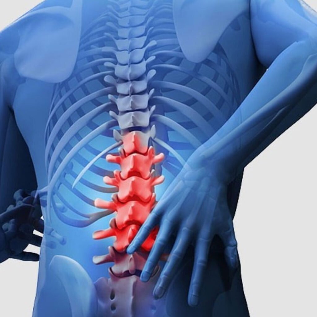
Lumbar spondylosis is a degenerative condition affecting the lower spine, commonly caused by aging, wear and tear, or injury. It involves the breakdown of spinal discs, joints, and ligaments, leading to stiffness, pain, and reduced flexibility in the lower back. In severe cases, it may compress nearby nerves, causing numbness or weakness in the legs.
Intervertebral discs lose flexibility and hydration, reducing cushioning between vertebrae and causing persistent lower back pain.
Facet joints wear out, leading to stiffness, inflammation, and discomfort, especially during movement or prolonged activity.
The narrowness of the spinal canal compresses the veins, resulting in back pain, tingling, numbness and leg weakness.
One vertebra slips forward over another, causing nerve compression, lower back pain, and walking difficulties.
Continuous dull or sharp lower back pain worsens with physical activity and prolonged sitting.
Morning stiffness limits spinal movements, making bending, twisting, and standing extremely uncomfortable.
Compressed veins cause tingling, numbness and weakness in the legs, sometimes affecting the ability to walk normal.
Low spinal flexibility makes it difficult to perform daily tasks and physical activities.
The lower back pain is reduced due to nerve root compression.
MRI scans offer spinal discs, nerves and softened tissues around the nerves. This disk helps in detecting herniation, nervous compression and degeneration, which provides a clear understanding of the lumbar spondylosis.
CT scans produce extremely wide cross-depleted images of the spine. They are especially useful in identifying bony changes, spinal canal narrowness and structural deformity, when X-rays provide better insight when they fail to give adequate information.
Milography involves injecting a contrast dye in the spinal canal, showing nerve roots and spinal structures on the imaging scan. It is used when MRI or CT results are individual, especially in spondylosis cases of complex lumbar.
EMG measures muscle electrical activity to identify abnormal muscle reactions caused by nerve compression. This probe helps evaluate the degree of nerve damage and effectively separates spondylosis of lumbar from other neuromuscular disorders.
The NCS measures the speed and strength of the electrical signals traveling through peripheral veins. It identifies damage caused by nervous compression, obstructions, or lumbar spondylosis, often performed with EMG for a broad neurological evaluation.
A bone density test assesses bone strength and detects osteoporosis, which may spoil spondylosis of the lumbar. Weak bones increase spinal degeneration risk, making this test important to patients, especially older adults or people with recurrent fractures.
Blood tests help control infection, inflammatory conditions, or autoimmune diseases that can mimic spondylosis symptoms of lumbar. They are especially useful in separating spondylosis from arthritis, effectively, ankyloging spondylitis, or other inflammatory spinal disorders.
Discography involves injecting a contrast dye in the spinal disc to identify damaged or impure discs. This is done when other imaging methods cannot detect the exact source of pain, providing clinical information targeted for treatment.
A wide physical examination assesses asana, spinal flexibility, muscle strength, reflexes and nerve function. Doctors conduct agitation tests to evaluate pain trigger and neurological deficit, which helps determine the severity and progress of spondylosis of the lumbar spondylosis.
Copyright © 2025 – Designed by Camsol Advertising