Peptic ulcer disease is a condition in which open lesions are called ulcers, on the internal lining of the abdomen, the upper intestine (duodenum), or on the lower esophagus. These ulcers develop due to imbalance between stomach acid and protective mechanisms, often due to factors such as helicobacter pylori infection, NSAID, smoking, alcohol, or excessive use of stress. Common symptoms include abdominal pain, swelling, resentment, nausea and indigestion.
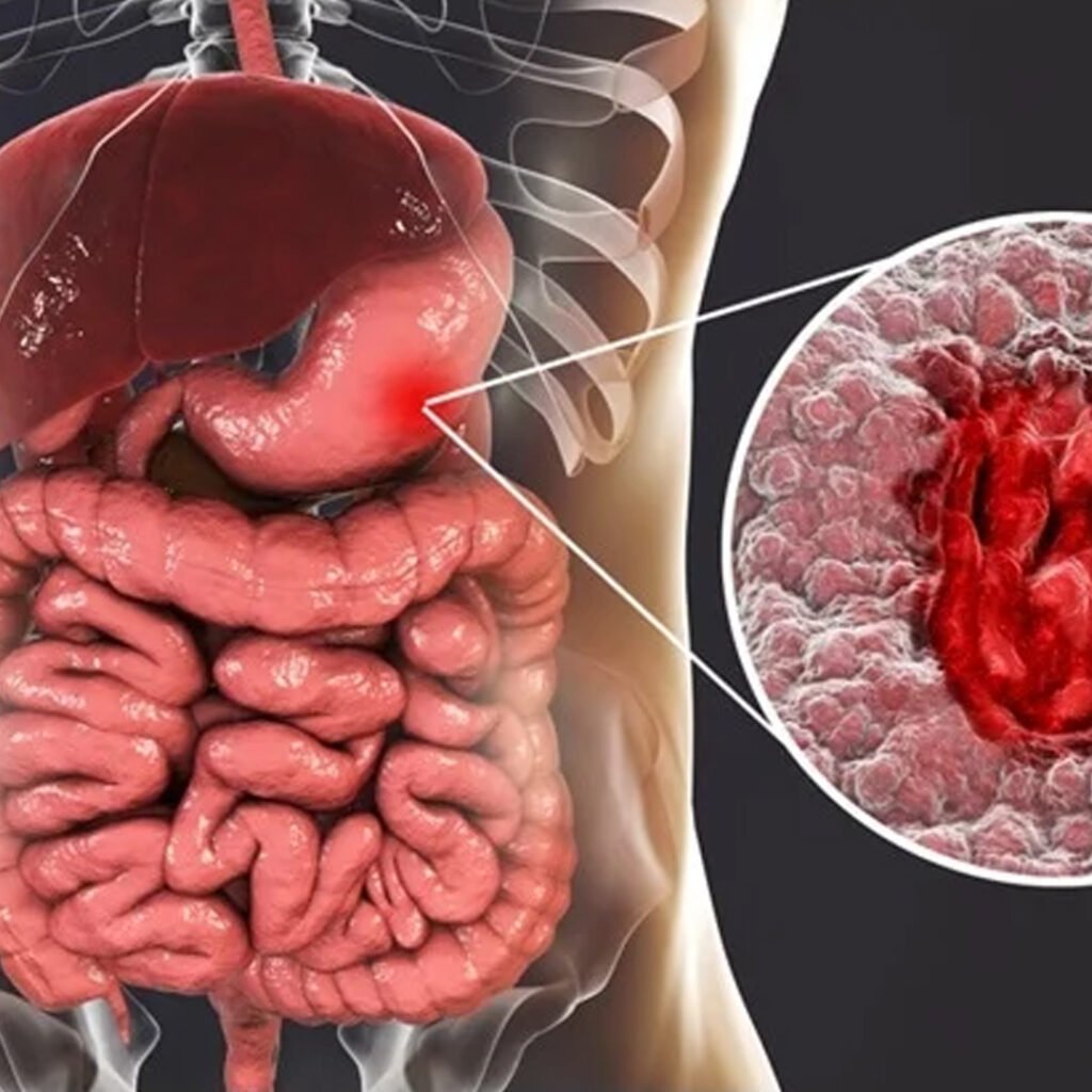
Gastric ulcers are formed on the internal lining of the abdomen, mainly H. Due to pylori infection, NSAID use, or increase in acid production.
Duodenal ulcers appear in the upper small intestine, commonly linked with H. pylori infection and excessive stomach acid secretion.
Esophageal ulcers develop in the lower esophagus, often caused by chronic acid reflux, GERD, or frequent vomiting episodes.
Stress ulcers occur during severe illness, trauma, burns, or surgery due to decreased blood flow and excessive stomach acid production.
The upper abdomen causes irritation or pain pain, usually deteriorating on an empty stomach.
Patients experience swelling, discomfort and perfection after eating small amounts of food.
Ulcers may cause nausea and sometimes vomiting, especially after heavy or acidic meals.
Frequent heartburn occurs due to acid reflux, causing burning sensations in the chest area.
Severe ulcers may lead to vomiting blood or passing black, tarry stools indicating bleeding.
This non-invasive test detects active Helicobacter pylori infection by measuring exhaleed carbon dioxide after swallowing a urea solution. It is used to confirm the participation of bacteria in highly accurate and widely peptic ulcer disease diagnosis.
This test confirms the presence of infection, H in stool samples. Pylori identifies antigen. It is simple, non-invasive and useful for both early diagnosis and monitoring of the effectiveness of treatment in peptic ulcer disease.
A CBC chronic ulcer helps to detect anemia as a result of bleeding. Low hemoglobin levels or low hematocrit values indicate potential gastrointestinal blood loss, helping to evaluate possible complications such as ulcers’ severity and hidden bleeding.
These tests measure pancreatic enzyme levels to control pancreatitis, which can mimic the symptoms of ulcers. The level of elevated enzyme suggests pancreatic involvement or complications associated with severe peptic ulcer disease cases.
Patients have to swallow a barium solution, and X-ray imaging exposes the abdomen and duodenum. This test ulcers identify crater, gastric outlet barrier and other structural abnormalities when endoscopy is not easily available.
These tests measure the amount of acid produced by the stomach. Unusually high acid levels indicate hypersacresly states such as Zollinger-Elison syndrome, which increases ulcer risk and severity in older cases.
These tests evaluate liver and kidney health, ensuring safe use of ulcer medications. Impaired function can worsen symptoms or limit treatment options, especially in patients with chronic peptic ulcer disease complications.
Ultrasound detects complications like gastric outlet obstruction, perforation, or swelling around the stomach. It’s non-invasive and useful when ulcers cause associated fluid accumulation or surrounding tissue inflammation in chronic conditions.
A CT scan provides detailed imaging of the stomach and nearby structures, identifying perforations, obstructions, or suspected malignancies. It’s particularly valuable in complicated or non-healing chronic peptic ulcer disease cases.
Copyright © 2025 – Designed by Camsol Advertising