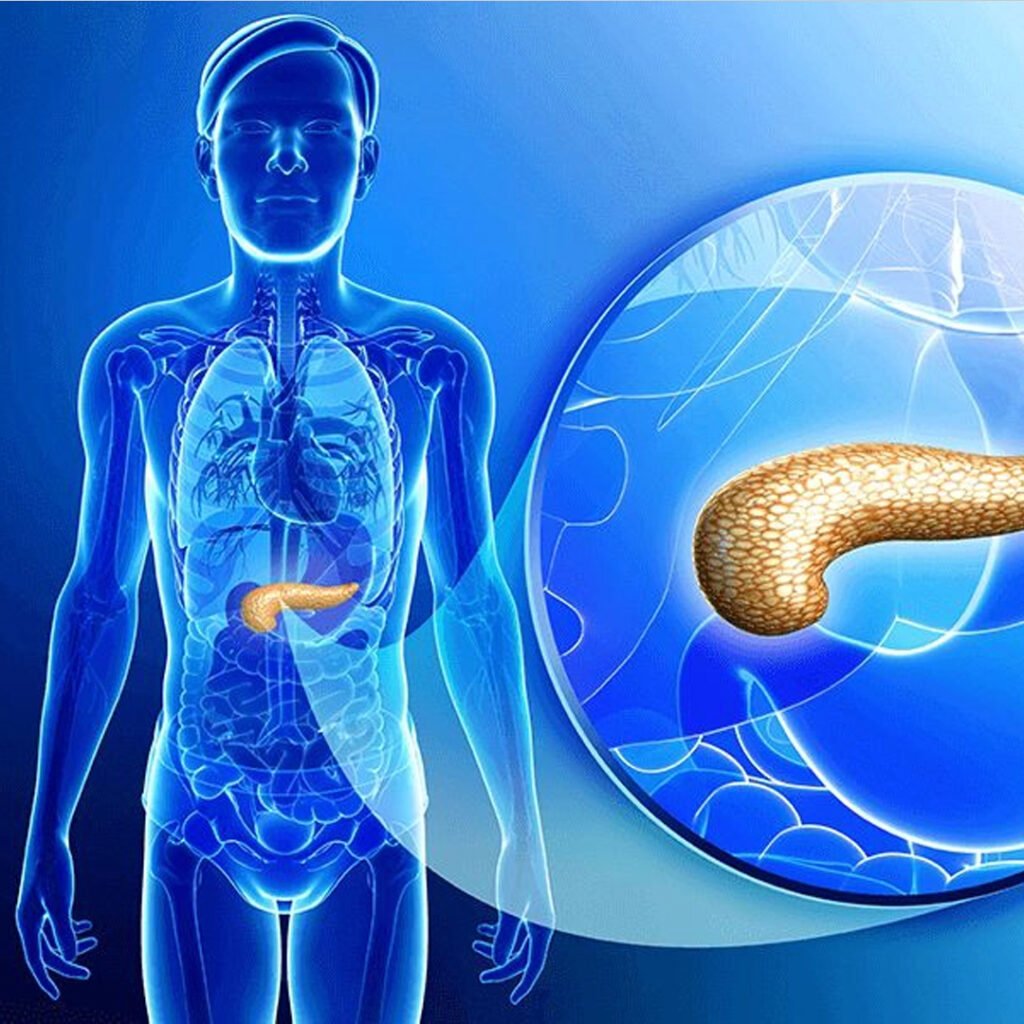
Acute pancreatitis is a sudden inflammation of the pancreas, an important organ responsible for the production of digestive enzymes and regulating blood sugar levels. This condition occurs when the digestive enzymes become active inside the pancreas instead of the small intestine, causing irritation and tissue damage. This can range from mild discomfort to serious, life-threatening disease. Common causes include bile stones, excessive alcohol consumption, certain medications, infections and stomach injuries. Initial diagnosis and treatment are necessary to prevent complications such as organ failure, infection and chronic pancreatitis.
Light acute pancreatitis causes limited pancreatic inflammation without organ failure and usually resolves quickly with conservative treatment and accessory medical care.
Minor severe acute pancreatitis involves transient organ dysfunction or local complications, usually improved within a week with timely supporting management.
Severe acute pancreatitis causes widespread pancreatic necrosis and frequent organ failure, requiring intensive medical care and sometimes surgical intervention for recovery.
Necrotizing acute pancreatitis occurs when pancreatic tissue dies due to severe inflammation, often leading to infection and life-threatening complications.
Suddenly, acute upper abdominal pain reaches the back, deteriorating after food or alcohol.
Frequent nausea with frequent vomiting is usually seen in cases of acute pancreatitis.
The upper abdomen gets tender to touch, often associated with muscle guarding and inflammation.
Infections and inflammation rapidly raise body temperature with weak pulse.
The obstruction of the bile ducts caused by the yellow skin and the tears of the eyes due to bile stones.
Severe upper abdominal pain reaches the back, deteriorating after meals.
Serum lipase is a pancreatic enzyme more specific than amylase. Levels rise within hours of pancreatic injury and remain elevated longer. Measuring lipase helps confirm acute pancreatitis and assess severity. Persistent elevation can indicate ongoing pancreatic damage or complications, making it a reliable marker for diagnosis and monitoring.
CBC evaluates white blood cells, hemoglobin, and platelets. Leukocytosis indicates inflammation or infection, while low hemoglobin suggests bleeding. Platelet counts can reflect systemic stress or sepsis. CBC provides information on the body’s inflammatory response and helps identify complications such as anemia, infection, or systemic involvement in acute pancreatitis patients.
LFTs measure bilirubin, ALT, AST, and alkaline phosphatase levels. Abnormalities may indicate gallstones, bile duct obstruction, or liver involvement. Elevated bilirubin suggests obstructive pancreatitis. Monitoring LFTs helps determine the cause of pancreatitis, guides management, and identifies concurrent liver complications that require timely intervention.
Electrolytes such as sodium, potassium, calcium and chloride are monitored during pancreatitis. Vomiting, loss of fluid, and inflammation can cause imbalance, causing heart, neuromuscular, or kidney complications. Curing these imbalances is necessary for the stability of the patient and prevent systemic effects associated with acute pancreatitis.
Pancreatic inflammation can deteriorate insulin secretion, resulting in hyperglycemia. It is important to monitor blood sugar levels to prevent metabolic complications and manage effectively acute pancreatitis. Constant high glucose can indicate endocrity of endocrine pancreatic and require temporary insulin therapy or careful monitoring during recovery.
Abdominal ultrasound is a non-invasive imaging technique used to evaluate the pancreas, detect gallstones and assess ductal obstruction. It also helps in identifying pancreatic inflammation or fluid collection. Ultrasound is often the first imaging study for suspected pancreatitis because it is safe, widely available, and does not include radiation risk.
CECT provides detailed imaging of the pancreas, helps in detecting necrosis, fluid collection, prochemist or other complications. It is particularly useful in severe cases to assess the limit and severity of the disease. Time is important, usually the symptom is done 48–72 hours after the onset to obtain optimal clinical information.
MRCP is a non-invasive imaging technique that imagines pancreatic and bile ducts. It detects stones, strictness, or ductal anomalies, causing acute pancreatitis. MRCP avoids radiation exposure and is useful for interfering or planning surgery, especially in patients where ultrasound or CT conclusions are indiscriminate.
The EUS combines endoscopy and ultrasound to provide high-resolution images of the pancreas. It is highly sensitive to detect small stones, ulcers, tumors or structural abnormalities. The EUS can guide therapeutic interventions such as stone removal or biopsy, making it a valuable tool in the management of matters of complex or recurrent acute pancreatitis.
Copyright © 2025 – Designed by Camsol Advertising