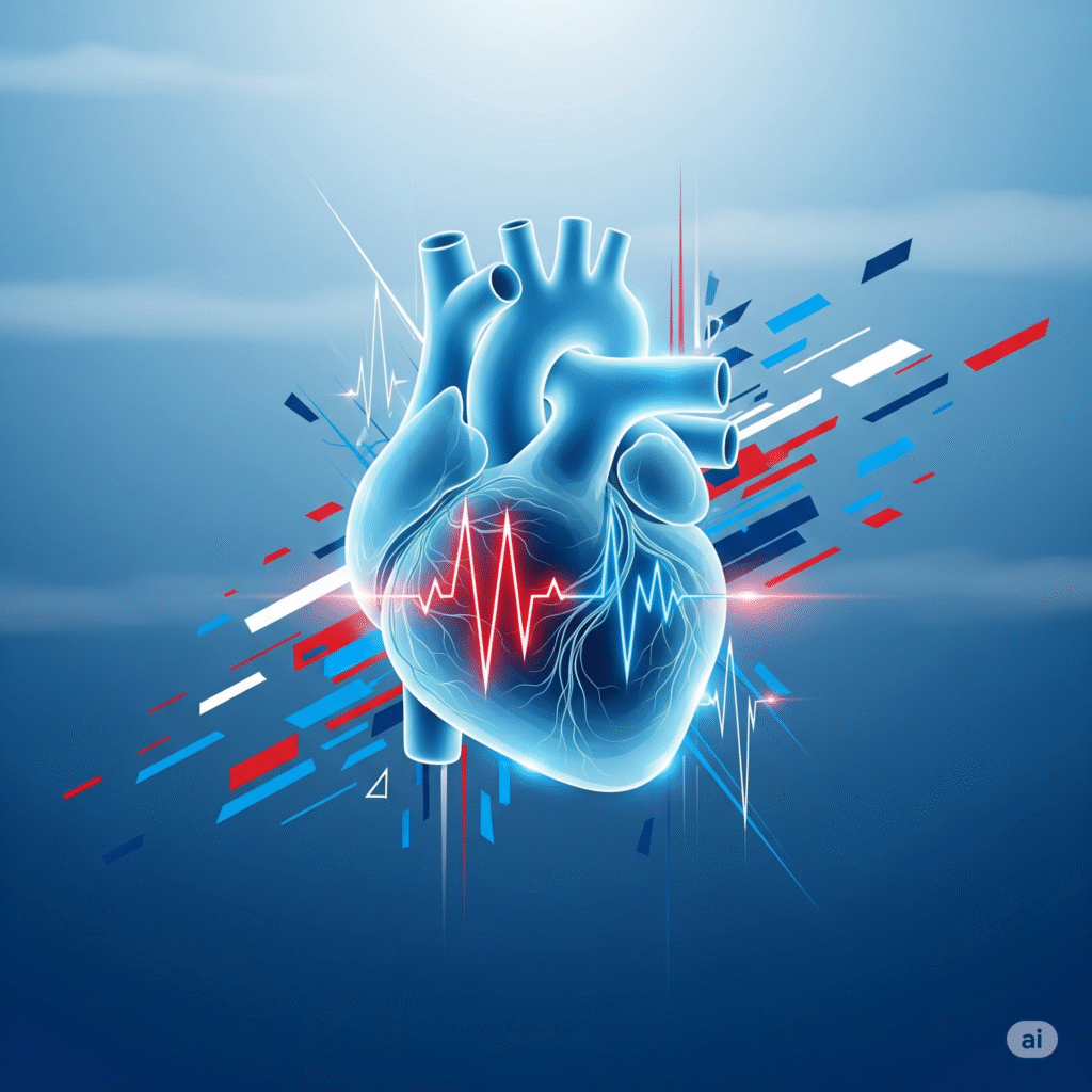
Cardiac arrhythmias are abnormal heart rhythms caused by disturbances in the heart’s electrical system. These irregular rhythms can make the heart beat too fast (tachycardia), too slow (bradycardia), or erratically. Arrhythmias may be harmless, but some can be serious or even life-threatening, affecting the heart’s ability to pump blood efficiently.
Rapid, irregular electrical signals in the atria cause an uncoordinated, fast heartbeat.
A slow heart rate, usually below 60 beats per minute, often due to problems in the sinus node.
Fast heart rhythm originating from the ventricles that may impair blood flow and lead to cardiac arrest.
Abnormally fast heart rhythm starting above the ventricles, often due to abnormal electrical pathways
Rapid, flutter, or irregular heartbeat sensation in chest.
due to a decrease in blood flow to the brain due to irregular heartbeat.
Disabled heart pumping supplies insufficient oxygen to the lungs and the body.
occurs when arrhythmia affects blood supply to the heart muscle.
A sudden loss of consciousness due to blood pressure or fall in heart production.
A portable ECG device worn for 24–48 hours to monitor irregular heartbeats.
Worn for weeks; records heart rhythm during symptoms.
Uses ultrasound to assess heart structure and function.
Invasive test to map electrical signals and locate arrhythmia origin.
Monitors heart rhythm during physical exertion to provoke arrhythmias.
Consider with obstructive urinary symptoms, persistent haematuria, family history of polycystic kidney disease or progressive CKD. Small kidneys suggest chronicity. Asymmetric renal size suggests renovascular or congenital disease
Provides detailed images of heart structures and tissues.
Copyright © 2025 – Designed by Camsol Advertising