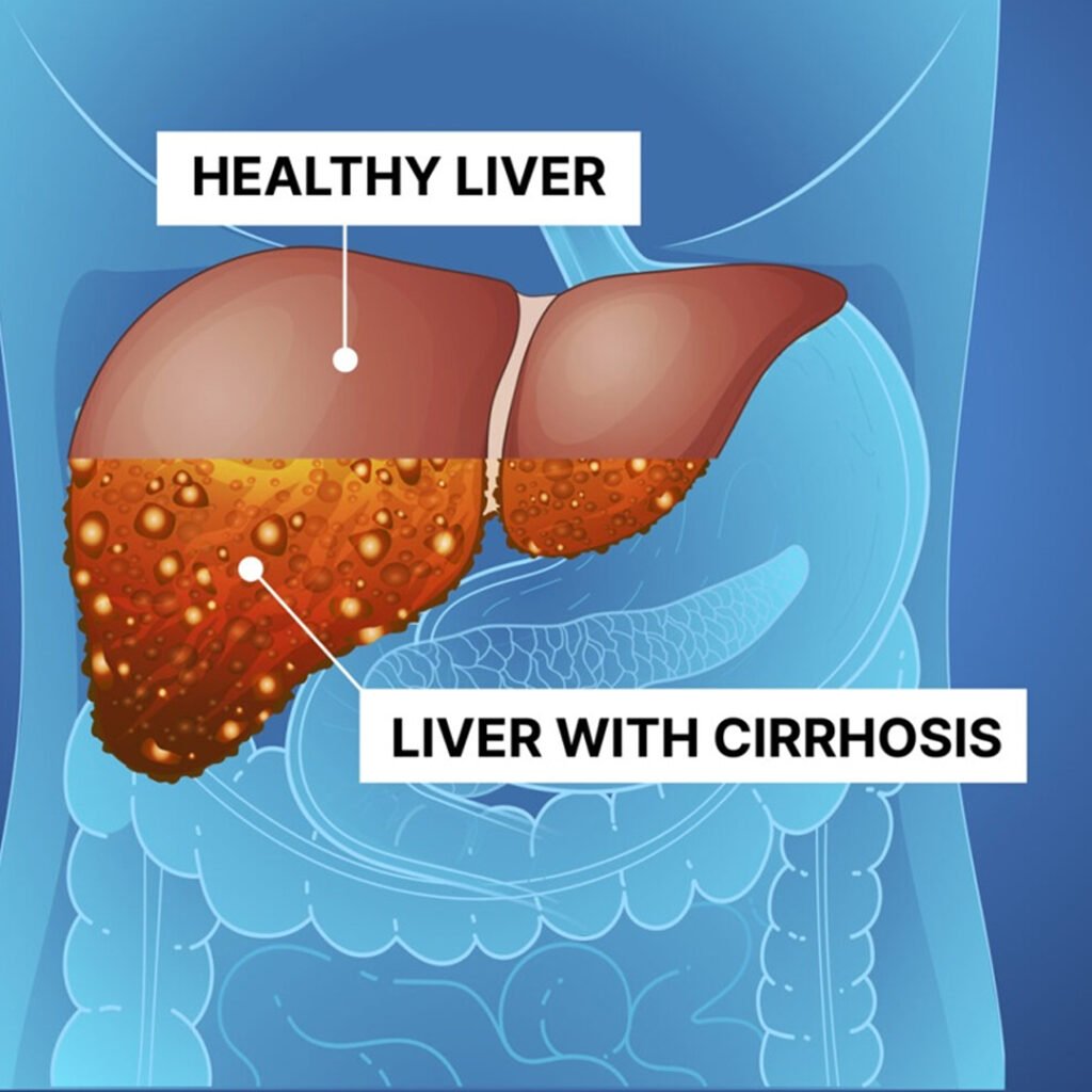
Cirrhosis is a chronic, progressive liver disease characterized by scarring and impaired liver function. It results from long-term damage caused by factors like alcohol, viral hepatitis, or autoimmune diseases. Over time, normal liver tissue is replaced by fibrous tissue, reducing liver efficiency and potentially leading to liver failure, complications, or portal hypertension.
Caused by prolonged alcohol abuse, leading to liver scarring and impaired function.
Develops due to chronic viral hepatitis B or C infection damaging liver tissue.
Results from long-term bile flow obstruction, causing bile accumulation and liver fibrosis.
Follows massive liver cell death due to toxins, drugs, or severe infections.
Yellowing of skin and eyes occurs due to bilirubin accumulation from liver dysfunction.
Persistent tiredness develops because the damaged liver cannot efficiently metabolize nutrients and toxins.
Fluid accumulates in the abdomen due to portal hypertension and reduced liver protein production.
Reduced clotting factor synthesis by the liver leads to frequent bruising and bleeding.
Enlarged liver and spleen result from chronic inflammation and portal hypertension in cirrhosis.
Assesses red and white blood cells, hemoglobin, and platelets. Cirrhosis often causes anemia, leukopenia, or thrombocytopenia due to portal hypertension, hypersplenism, and bone marrow suppression.
Non-invasive imaging used to evaluate liver size, texture, nodules, and fatty changes. Detects early cirrhotic changes, ascites, and spleen enlargement for monitoring disease progression.
Provides detailed cross-sectional images of the liver, identifying fibrosis, regenerative nodules, hepatomegaly, and complications like portal vein thrombosis or ascites.
High-resolution imaging visualizes liver architecture, detects fibrosis, nodules, and vascular abnormalities. Helps assess disease severity and differentiate benign versus malignant lesions in cirrhosis.
Evaluates clotting ability, which is reduced in cirrhosis due to impaired synthesis of clotting factors. Prolonged PT indicates advanced liver dysfunction and bleeding risk.
Measures bilirubin levels to assess liver’s excretory function. Elevated bilirubin causes jaundice and indicates impaired bile processing or hepatocellular damage.
Monitors for hepatocellular carcinoma development, which is a common complication in patients with long-standing cirrhosis. High AFP levels suggest malignancy.
A sample of liver tissue is examined microscopically to confirm cirrhosis, assess fibrosis stage, and identify underlying etiology, such as alcohol, hepatitis, or autoimmune disease.
Detects esophageal and gastric varices caused by portal hypertension. Helps prevent life-threatening bleeding by guiding prophylactic treatment like banding or medication.
Copyright © 2025 – Designed by Camsol Advertising