Gallstones are solid, stone-like deposits that develop in the gallbladder, a small organ located under the liver that stores and releases bile. They form when there is an imbalance in bile components, such as cholesterol, bile salts, or bilirubin, causing crystallization. Gallstones can range in size from tiny grains to large stones and may remain asymptomatic for years or cause severe pain and digestive issues when they block bile flow, leading to complications like inflammation, infection, or jaundice.
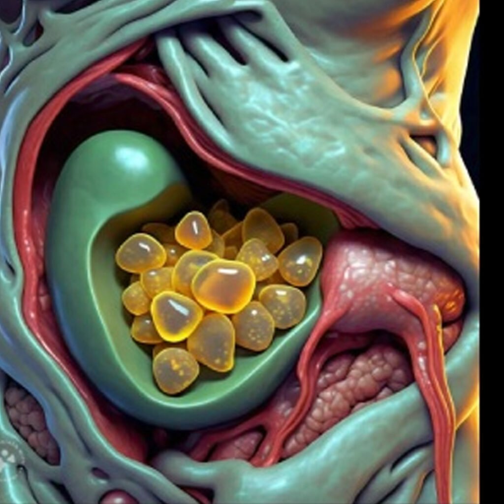
These are the most common type, formed primarily from hardened cholesterol in bile. They can develop slowly and may remain asymptomatic for a long time.
Dark-colored stones that form due to excess bilirubin, often linked to liver disease, infections, or certain blood disorders.
These stones are composed of a combination of cholesterol, calcium salts, and bilirubin, representing features of both cholesterol and pigment stones.
Uncommon gallstones caused by genetic factors, bile duct infections, or metabolic disorders, and may require special medical attention.
Gallstones often cause sharp or cramping pain in the upper right abdomen, especially after fatty meals.
Gallstones can irritate the digestive system, leading to frequent nausea and occasional vomiting episodes.
Patients may experience discomfort, bloating, and indigestion after eating heavy or oily foods.
If a gallstone blocks the bile duct, yellowing of the skin and eyes (jaundice) can occur.
Infection caused by trapped gallstones may lead to fever, chills, and general weakness.
LFTs help assess liver health and detect bile flow obstruction caused by gallstones. Elevated bilirubin, alkaline phosphatase, and liver enzymes indicate gallstone-induced blockage or inflammation. These tests are often performed alongside imaging to evaluate the extent of liver and biliary system involvement.
EUS combines endoscopy and ultrasound for highly detailed images of the bile ducts, pancreas, and gallbladder. It is useful for detecting small gallstones and sludge not visible on standard ultrasound. This procedure is minimally invasive and particularly beneficial for complex cases involving bile duct stones.
MRCP uses MRI technology to visualize the biliary and pancreatic ducts in detail. It is a non-invasive test that helps identify gallstones in the bile ducts, strictures, and other abnormalities. MRCP is particularly useful when ultrasound results are inconclusive or when bile duct stones are suspected.
ERCP is both a diagnostic and therapeutic tool. It visualizes the bile ducts using contrast dye and X-ray, detects gallstones, and allows their removal if present. It is mainly used when bile duct obstruction or infection is suspected, but it carries some risks, including pancreatitis.
A CT scan provides detailed cross-sectional images of the abdomen and detects gallstones, gallbladder inflammation, or complications like perforation or abscess. While less sensitive than ultrasound for gallstones, it is useful in identifying other abdominal conditions that may mimic gallstone symptoms.
A HIDA scan evaluates gallbladder function and bile flow. It involves injecting a radioactive tracer and taking images to track bile movement. It is particularly helpful in diagnosing acute cholecystitis, where gallstones block bile flow, and can assess gallbladder ejection fraction in chronic cases.
A CBC helps detect signs of infection or inflammation caused by gallstones. Elevated white blood cell counts suggest acute cholecystitis or gallbladder infection. This test, combined with imaging and liver function tests, provides valuable insights into complications associated with gallstones.
These enzymes are tested when gallstones are suspected to cause pancreatitis. Elevated levels indicate gallstone-induced blockage of the pancreatic duct, leading to inflammation. Measuring these enzymes is crucial for detecting life-threatening complications and guiding appropriate treatment strategies.
PTC involves injecting contrast dye directly into the bile ducts through the liver and capturing X-ray images. It is primarily used when ERCP is not possible or fails. This test helps identify gallstones, strictures, and blockages in the bile ducts and assists in planning treatment.
Copyright © 2025 – Designed by Camsol Advertising